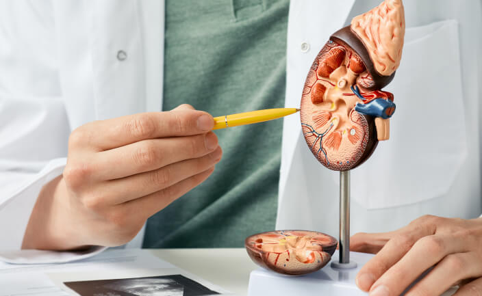
Common Symptoms of Kidney Stones
Common indicators that necessitate a medical evaluation include:
- Severe back or side pain, often referred to as flank pain. This pain is often intermittent and intense, changing location as the stone moves.
- The presence of blood in the urine, known as haematuria.
- Painful or frequent urination, sometimes signalling a developing infection.
- Nausea or vomiting, often associated with the intensity of the pain.
- Fever and chills, which suggest a serious complication like an infected, obstructed kidney (pyelonephritis or urosepsis), requiring immediate attention.
What Are the Causes of Kidney Stones?
Kidney stones develop when urine contains high concentrations of substances such as calcium, oxalate, or uric acid, without enough fluid to keep them dissolved. Stones may also form when natural inhibitors that prevent crystals from sticking together are lacking. Understanding these contributing factors is an important part of the diagnostic process, as long-term prevention depends on identifying the stone type.
There are four main categories of kidney stones:
1. Calcium Stones
The most common type, calcium stones usually form as calcium oxalate. Their development may be linked to dietary patterns, metabolic conditions, or high doses of vitamin D.
2. Struvite Stones
Often associated with urinary tract infections (UTIs), struvite stones can enlarge rapidly and become sizable, which may necessitate surgical removal.
3. Uric Acid Stones
Uric acid stones are more likely in people with low fluid intake, excessive fluid loss, or conditions such as gout. They may also develop during chemotherapy. These stones form when urine becomes highly concentrated or acidic.
4. Cystine Stones
Cystine stones are rare and occur in individuals with cystinuria, a hereditary condition that causes the kidneys to excrete large amounts of cystine. Because this amino acid dissolves poorly in urine, stone formation becomes more likely.
How Are Kidney Stones Diagnosed?
Suspected kidney stones are assessed using a systematic approach that confirms their presence, evaluates their size and location, and determines any effect on kidney function. Diagnosis generally combines laboratory testing with imaging studies, including:
Urine Test (Urinalysis)
Urinalysis helps detect blood, signs of infection and elevated levels of stone-forming substances such as calcium or uric acid. These findings offer valuable clues about the stone type and highlight any associated complications.
Blood Tests
Blood tests are carried out to evaluate kidney function (by measuring creatinine and urea levels) and to check for high levels of calcium or uric acid in the blood, which can point toward an underlying cause.
Imaging Studies
Imaging is essential to pinpoint the exact location, size, and number of stones.
- Computed Tomography (CT) Scan: A non-contrast CT scan is considered the gold standard for kidney stone diagnosis. It can detect almost all stone types, accurately measure size and density, and identify stones that may not appear on standard X-rays.
- Ultrasound: Ultrasound is a quick, non-invasive option and is often used as an initial test, particularly for pregnant patients or those who need to avoid radiation. It can identify kidney swelling caused by obstruction and detect many larger stones.
- X-rays (Kidneys, Ureters, Bladder or KUB): X-rays of the kidneys, ureters and bladder can detect radiopaque stones, mainly those containing calcium. However, some stone types may not be visible using this method alone.
How Are Kidney Stones Treated?
Once a kidney stone has been identified, treatment is tailored to its size, location and composition, as well as your overall health and symptoms. Management can range from careful monitoring to active removal, depending on how likely the stone is to pass naturally.
Smaller stones may be managed conservatively with increased fluid intake and medication to relieve pain, alongside regular follow-up to ensure they are progressing safely. Larger stones, or those causing obstruction, infection or ongoing discomfort, usually require more active intervention, such as:
Extracorporeal Shockwave Lithotripsy (ESWL)
A non-invasive option, ESWL uses focused sound waves to break stones into small fragments that can pass naturally through the urine. It is most suitable for stones located in the kidney or upper ureter.
Ureteroscopy (URS)
During this procedure, a thin flexible scope is guided through the urethra and bladder into the ureter or kidney. In some cases, a laser is used to fragment the stone, after which the pieces are removed or left to pass on their own.
Percutaneous Nephrolithotomy (PCNL)
For very large or complex stones, PCNL provides a direct approach. A small incision is made in the back to allow instruments to enter the kidney and remove the stone.
Laparoscopic or Robotic Stone Surgery
When stones are very large or cannot be removed with simpler procedures, surgeons may use laparoscopic or robotic techniques. These methods are occasionally chosen for complex stones, such as large staghorn formations, that need to be fully removed.
Finding Relief and Preventing Recurrence
The diagnostic process for kidney stones follows a structured approach that brings together symptom assessment, laboratory tests, and appropriate imaging. This combination helps clinicians determine the stone’s size, location, and impact on the urinary system. With this information, the most suitable treatment can be selected to manage the condition effectively and support the patient’s long-term kidney health.
If you are experiencing symptoms or have concerns about kidney stones, seeking early specialist assessment can provide clarity and timely relief. Contact our clinic today to arrange a consultation with a kidney stone specialist and take a confident step towards effective treatment and peace of mind.